Broca and Wernicke Areas
Broca’s area and Wernicke’s area are two important regions in the brain that are involved in language processing.
Broca’s area, located in the frontal lobe of the left hemisphere, is primarily responsible for speech production and fluent language expression. It plays a crucial role in coordinating the movements of the speech muscles and generating grammatically correct sentences. Damage to Broca’s area can result in a condition called Broca’s aphasia, characterized by difficulty in speaking fluently while comprehension remains relatively intact.
On the other hand, Wernicke’s area, situated in the temporal lobe of the left hemisphere, is responsible for language comprehension. It helps in making sense of spoken and written words and enables the understanding of meaning and context. When Wernicke’s area is damaged, a person may exhibit Wernicke’s aphasia, wherein they may produce fluent but nonsensical speech and have difficulty understanding language.
Primary motor cortex
The primary motor cortex, located in the frontal lobe of the brain, plays a crucial role in the control of voluntary movements. Here are some key facts about the primary motor cortex:
- Location: The primary motor cortex is situated just in front of the central sulcus, a prominent groove that separates the frontal and parietal lobes of the brain.
- Motor Control: This region is responsible for planning, executing, and controlling voluntary movements of the body. It sends signals to different parts of the body, enabling smooth and coordinated motion.
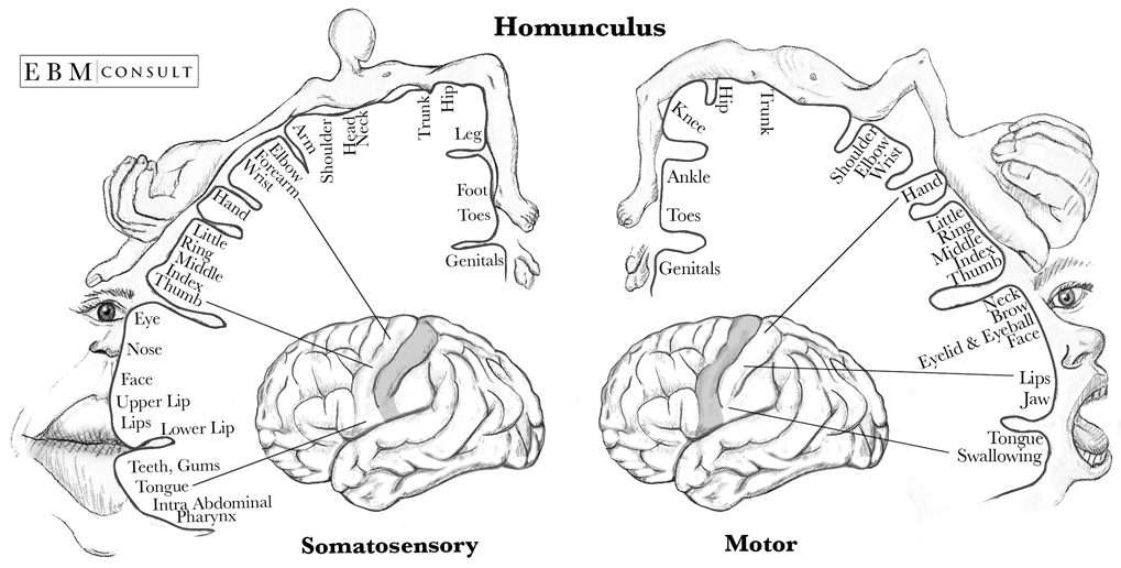
- Topographical Organization: The primary motor cortex exhibits a somatotopic organization, meaning that different body parts are represented in an orderly manner. The amount of space dedicated to a specific body part is proportional to its level of fine motor control. For example, the area dedicated to the fingers is relatively larger compared to the area representing the trunk.
- Motor Homunculus: The somatotopic arrangement in the primary motor cortex is often depicted as a “motor homunculus,” a distorted representation of the human body. It visually represents the disproportionate allocation of cortical space to different body parts based on their fine motor control requirements.
- Motor Learning and Plasticity: The primary motor cortex is involved in motor learning and can undergo structural and functional changes, known as plasticity. Through practice and repetition, the primary motor cortex adapts and refines its connections to improve motor skills.
- Connection with other Brain Areas: The primary motor cortex collaborates closely with other brain regions, such as the supplementary motor area and the basal ganglia, to coordinate complex movements and maintain motor balance.
Understanding the primary motor cortex enhances our knowledge of how the brain controls movement and helps in the diagnosis and treatment of various motor disorders.
Case Study – Phinehas Gage (1848)
The case study of Phinehas Gage in 1848 is one of the most renowned and intriguing cases in the field of neuroscience. Phinehas Gage was a railroad construction worker who survived a severe brain injury that dramatically altered his personality and behavior.
During a work accident, Phinehas’ skull was penetrated by a large iron rod, which entered through his cheek and exited through the top of his head. Miraculously, he survived the incident, but his once calm and responsible demeanor was replaced by impulsivity and unrestrained behavior.
The observation of Phinehas Gage’s post-injury transformation provided valuable insights into the localization of brain function. Before the accident, he was considered hardworking and reliable, but afterwards, he became impulsive, unreliable, and socially inappropriate.
This case study played a significant role in shaping our understanding of the frontal lobe’s importance in personality and behavior regulation. Phinehas Gage’s experience highlights the intricate relationship between brain structures and their influence on human behavior, paving the way for further research in the field of neuroscience.
Although the case of Phinehas Gage occurred over 170 years ago, it continues to captivate researchers, medical professionals, and the general public, as it offers a compelling glimpse into the complexities of the human brain.
The brainstem
There are three parts in the brainstem – the midbrain, pons and medulla oblongata.
- The medulla: contains several centers responsible for vital autonomic functions like digestion, breathing, and the control of heart rate.
- The pons: crucial for conveying information about movement from the cerebral hemispheres to the cerebellum.
- The midbrain: controls many sensory and motor functions, including eye movement and the coordination of visual and auditory reflexes.
They lie above the midbrain and is made up of two major structures:
Thalamus: processes most of the information that reach the cerebral cortex. hypothalamus: regulates autonomic, endocrine and visceral functions
The cerebellum
Apart from its role in motor control, the cerebellum also plays a crucial role in coordination, balance, and posture. It receives sensory information from various parts of the body and integrates it with motor signals from the brain to ensure smooth and precise movement. Additionally, the cerebellum has been implicated in cognitive functions such as attention, language, and problem-solving. Its intricate structure and intricate network of connections make the cerebellum an essential component of our nervous system.

Leave a comment